What To Order And When: An Overview Of Ordering Advanced Imaging For Musculoskeletal Conditions Specific To The Chiropractic Office

One of the most common questions I receive from clients and colleagues is always: “I have this clinical suspicion and/or x-rays finding…what advanced imaging do I order?” The goal of this article is to give basic guidelines regarding what specific advanced imaging should be ordered for the more commonly encountered MSK conditions in a chiropractic office. This article will not be an all-encompassing guide regarding all advanced imaging options for every possible condition, as that would be far too expansive to be useful. This article should be a quick and useful resource to help guide your decision making for your more commonly encountered conditions.
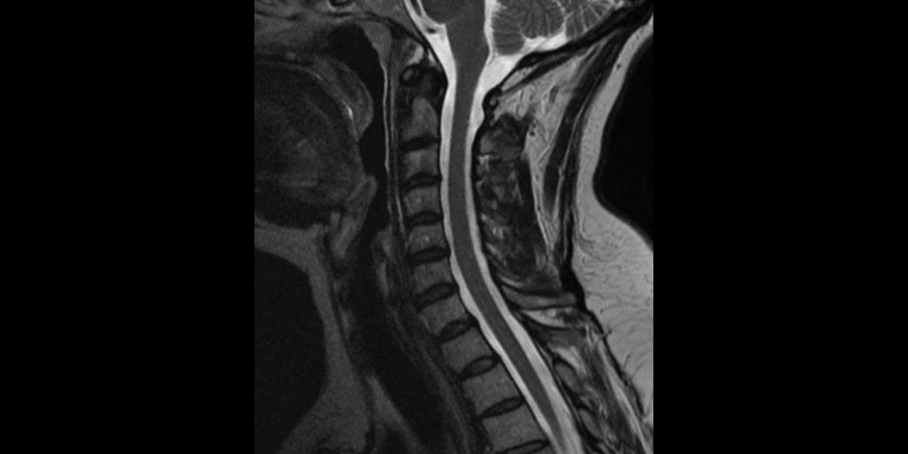
When considering advanced imaging for MSK conditions, MRI is most often the modality of choice for multiple reasons. Generally speaking, MRI can provide a more detailed image of soft tissue structures and marrow changes. Another very important advantage of MRI vs CT is that MRI does not use ionizing radiation. Of course, there are contraindications to MRI, which will be discussed at the conclusion of this article.
There are certain situations in which CT is superior to MRI, particularly in trauma cases. CT imaging is better at detecting subtle and complex fractures because MRI is not as sensitive in detecting subtle changes of cortical bone. Additionally, because CT is much faster than MRI, CT is preferred in emergency situations such as major trauma, acute head injury, or acute stroke.
Once the imaging modality has been determined, the next question is whether contrast is required. This article will focus on gadolinium contrast used in MRI. Iodinated contrast used in CT imaging plays an important role in imaging of the chest, abdomen/pelvis, and vasculature, but will not often be utilized in musculoskeletal imaging. When discussing contrast in MRI, we must first understand that there are three different types of contrast imaging in MRI: intravenous, intraarticular, and angiography. When we think of contrast, we generally think of intravenous contrast. Intravenous contrast (MRI with contrast) is when dye is injected into the patient’s vein and is absorbed into the patient’s tissues. Intraarticular contrast (MR arthrography) is injected directly into the joint capsule. MR angiography (MRA) is when the dye is injected intravenously but a specialized sequence is utilized to better analyze the vascular system. In general, most musculoskeletal conditions will not require contrast. Contrast is reserved for a clinical situation in which imaging/ clinical findings lead to a suspicion of malignancy or infection. Additionally, an important scenario in which MRI with and without contrast will be needed is in lumbar post-surgical patients (<10 years) to differentiate between re-herniation of the disc and post-surgical scar tissue (they can appear the same on a non-contrast study, but have very diffident clinical implications).
Table 1 is an imaging guideline reference sheet organized by body region, clinical findings, and procedure warranted.
Table 1- Contrast vs non-contrast guidelines
| Body region | Clinical indication | Procedure |
| Spine | Disc herniation Extremity pain/ weakness Radiculopathy Trauma Compression fracture Congenital anomalies Stenosis Spondylolysis | MRI without contrast |
| Spine | Discitis Osteomyelitis Tumor Post lumbar spinal surgery (<10 years) | MRI with and without contrast |
| Muscle/ tendon/ Soft tissue | Muscle tear Tendon tear/ tendinopathy | MRI without contrast |
| Muscle/ tendon/ Soft tissue | Cellulitis Abscess Morton’s neuroma Soft tissue tumor/mass Ulcer/ sinus tract | MRI with and without contrast |
| Bone (non-spinal) | Stress fracture Epiphyseal/ physeal injury Trauma (CT may be better if complex) | MRI without contrast |
| Bone (non-spinal) | Osteomyelitis Tumor | MRI with and without contrast |
| Joint | Internal derangement Ligament tear Meniscal tear Internal derangement Ligament tear Meniscal tear Cartilage injury Avascular necrosis Osteochondritis dissecans | MRI without contrast |
| Joint | Shoulder instability with suspected labral tear Hip labral tear TFCC tear/ injury Ligament tears of the ankle/ wrist Post surgical shoulder/ knee | MR arthroscopy |
Important contraindications to note for MRI (1) are included in Table 2. CT without contrast does not have any true contraindications, but does carry the risk of radiation exposure to the patient. Iodinated contrast utilized in CT imaging may be contraindicated in patients with renal disease and those that are allergic to iodine. Gadolinium contrast utilized for MRI has been associated with a rare, but severe, side-effect known as nephrogenic systemic fibrosis (2). This condition is more commonly seen in those with severe renal disease than those with normal renal function. Patients with any known renal disease should be further evaluated before undergoing a gadolinium enhanced study.
Table 2- MRI Contraindications
| Absolute contraindications | · Cardiac implantable electronic device (currently conditional cardiac implantable electronic devices are widely available and would not be contraindicated) · Metallic intraocular foreign bodies (skull x-ray of any patient that has welded or has metal injury to face) · Implantable neurostimulation system · Cochlear implants/ ear implant · Drug infusion pumps · Catheters with metallic components · Metallic bullets/ shrapnel · Cerebral artery aneurysm clips · Magnetic dental implants · Tissue expander · Artificial limb · Hearing aid · Piercing |
| Relative contraindications · Further investigation will be needed to ensure safety/ determine if specific device is allowed. · 3T and higher scanners may have additional contraindications | · Coronary and peripheral artery stents · Programmable shunts · Airway stents or tracheostomy · IUD (acceptable on 1.5 tesla scanner. 3T and higher need further evaluation) · Ocular prosthesis · Surgical clips/ wires · Joint replacement/ prothesis · IVC filter · Harrington rods (ok on 1.5T) · Tattoo within 6 weeks of completion · Colonoscopy within last 8 weeks |
It is always important to discuss any potential contraindications with your patient before referring them for an MRI. It is also very important that the patient informs the facility of any contraindication so it can be determined if the study can be modified. Patients that are very claustrophobic may need to be referred for an open-MRI scanner. MRI has not been found to be harmful to a pregnant patient or fetus, but pregnant patients should inform the radiology staff and should scanned be on a 1.5T machine (3). Additionally, MRI with contrast is not contraindicated in a breast feeding patient (4).
References:
- Ghadimi M, Sapra A. Magnetic Resonance Imaging Contraindications. [Updated 2023 May 1]. In: StatPearls [Internet]. Treasure Island (FL): StatPearls Publishing; 2024 Jan. Available from: https://www.ncbi.nlm.nih.gov/books/NBK551669/
- Ramalho M, Ramalho J, Burke LM, Semelka RC. Gadolinium retention and toxicity—an update. Adv Chronic Kidney Dis. 2017;24(3):138–146.
- Mervak BM, Altun E, McGinty KA, Hyslop WB, Semelka RC, Burke LM. MRI in pregnancy: Indications and practical considerations. J Magn Reson Imaging. 2019 Mar;49(3):621-631.
- Newman J. Breastfeeding and radiologic procedures. Can Fam Physician. 2007 Apr;53(4):630-1.


