Normal Anatomic Variants of the Cervical Spine

Editor’s note: Dr. Ruof is presenting the webinar “The Pediatric Spine: Normal Development, Anatomic Variants, and Common Pathologies” on March 11, 2025. Register to attend here.
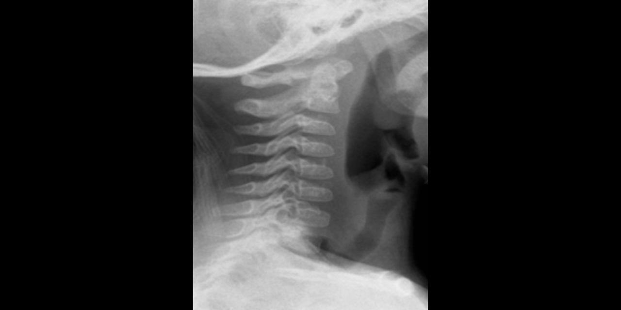
The pediatric cervical spine can be a source of confusion amongst practitioners, due to the anatomic variants that are normal findings. It is important for the clinician to understand and be able to recognize these variants in order to avoid unnecessary further imaging or unneeded intervention. This article will describe the common anatomic variants of the pediatric cervical spine, including 1) atlantodental interval widening, 2) pseudosubluxation of C2, 3) pseudo-Jefferson fracture, 4) wedging of the cervical vertebra, and 5) normal postural alterations.
1. Atlantodental interval widening
The atlantodental interval should be <3-mm in an adult, but it is important to remember that measurement up to 5-mm is considered normal in a child. This is due to the normal ligamentous laxity in children. Additionally, the pre-dental space is often V-shaped, a finding that should not be confused with ligamentous rupture.1


2. Pseudosubluxation of C2
Physiological anterior displacement of C2 on C3 is commonly seen in children, usually <7 years of age, less commonly in older children. It is most commonly seen at C2 on C3, but may less often be seen at C3 on C4. This finding is more pronounced in flexion and can present confusion in the presence of traumatic cervical injury .2 Utilization of Swischuk’s line is useful in differentiating pathological anterior cervical spine displacement from physiological displacement. This line is drawn from the anterior aspect of posterior arch of C1 to anterior aspect of posterior arch of C3 3 and should measure less than 2-mm. A measurement of greater than 2-mm indicates true subluxation. It is important to note that this line should only be used in the absence of other signs of instability and in the absence of prevertebral soft tissue swelling.


3. Pseudo-Jefferson’s fracture
Psuedo-Jefferson’s fracture or pseudospread of the atlas on the axis refers to the normal overhang of the lateral masses of C1 over the lateral edges of the body of C2. This is a normal physiologic finding in children up to the age of 7, most likely due to differential speed of growth of the atlas versus the axis.4 6-mm of overhang is the accepted maximal value of normal overhanging when adding the offset of the sides bilaterally.

4. Anterior wedging of C3 (or C4)
Anterior wedging of the C3 vertebral body, and less often, C4, is common in children up to the age of 12.5 Cervical vertebral bodies are oval in infancy and become more rectangular with age. However, in C3 and less often C4, wedging can persist. A hypothesis as to why this occurs in some children is that chronic, exaggerated hypermobility normally seen in childhood causes repetitive impaction of the vertebral body of C3 by C2, resulting in a subclinical insult that could impair normal ossification of cartilage at this site. The difference between anterior and posterior vertebral body height must be <3-mm to be considered normal.

5. Postural alterations of the cervical spine
Several postural alterations that may mimic muscle spasm and ligamentous injury are considered normal in children and include: non-uniform angulation between adjacent vertebra, absence of the normal lordotic curve, and absence of a flexion curvature seen on a lateral view with cervical flexion. These postural alterations may be seen up until the age of 16 and occur in 14-16% of the pediatric population.7
A solid understanding of these normal radiographic findings is imperative for any clinician treating children so that unnecessary imaging and treatment are avoided. In the absence of trauma, these findings are more easily interpreted as normal; however, confusion can occur when combined with trauma. Referral and advanced imaging are needed for any post-traumatic pediatric patients that are unable to communicate, have severe neck pain, or neurologic abnormalities.
References
- Bohrer SP, Klein A, Martin W 3rd. “V” shaped predens space. Skeletal Radiol. 1985;14(2):111-6.
- Ghanem I, El Hage S, Rachkidi R, Kharrat K, Dagher F, Kreichati G. Pediatric cervical spine instability. J Child Orthop. 2008 Mar;2(2):71-84.
- Swischuk LE. Anterior displacement of C2 in children: physiologic or pathologic. Radiology. 1977 Mar;122(3):759-63.
- Suss RA, Zimmerman RD, Leeds NE. Pseudospread of the atlas: false sign of Jefferson fracture in young children. (1983) AJR. American journal of roentgenology. 140 (6): 1079-82.
- Mortazavi, M., Gore, P.A., Chang, S. et al. Pediatric cervical spine injuries: a comprehensive review. Childs Nerv Syst 27, 705–717 (2011).
- Swischuk LE, Swischuk PN, John SD. Wedging of C-3 in infants and children: usually a normal finding and not a fracture. Radiology. 1993 Aug;188(2):523-6.
- Cattell, Hereward S.; Filtzer, DAVID L. Pseudosubluxation and other normal variations in the cervical spine in children: A study of one hundred and sixty children. The Journal of Bone & Joint Surgery 47(7):p 1295-1309, October 1965.


