Snapping Hip: The Iliopsoas Tendon

In addition to its contribution to various lumbopelvic problems, the iliopsoas tendon itself is a common source of hip pain and dysfunction. Potential problems can range from asymptomatic snapping to painful irritation of the tendon and underlying bursa.
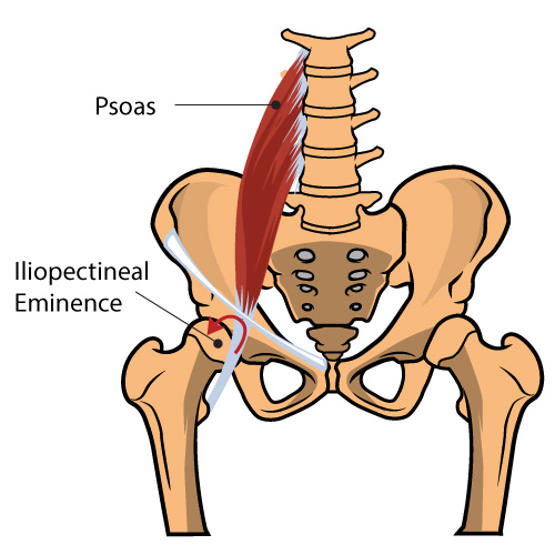
The deep fibers of the psoas muscle originate on the transverse processes of L1- L5, while the superficial fibers arise from the lateral surfaces of the lumbar vertebra and adjacent intervertebral discs. The iliacus muscle arises from the iliac fossa then converges with the psoas to form the iliopsoas tendon before inserting onto the lesser trochanter of the femur. The iliopsoas tendon travels across the anterior aspect of the acetabulum in a groove between the anterior inferior iliac spine (laterally) and the iliopectineal eminence (medially). The largest bursa in the body, the iliopsoas bursa, is positioned between the iliopsoas musculotendinous junction and the underlying bony pelvis. This bursa communicates with the hip joint in approximately 15% of adults. (1)
The powerful iliopsoas muscle is responsible for hip flexion and external rotation. When the muscle is excessively tight, its tendon may rub or even produce an audible snapping sound when passing over the underlying bony landmarks, including the iliopectineal eminence, lesser trochanteric bony ridge, anterior capsule of the femoral head, or anterior inferior iliac spine. (2-6) When painless, the condition is termed “asymptomatic internal snapping hip.” When accompanied by pain and/or dysfunction, the condition is known by a variety of terms, including: painful internal snapping hip, internal coxa saltans, iliopsoas tendinitis, iliopsoas tendinosis, iliopsoas tendinopathy, iliopsoas bursitis, or ilopsoas syndrome. The diagnoses “tendinitis” and “bursitis” are essentially synonymous, as inflammation in one inevitably generates inflammation in the neighboring counterpart, with a nearly identical presentation, evaluation, and management. (7)
In addition to “internal” snapping hip, clinicians should be aware of two other potential causes for reverberation: “external” snapping hip occurs when the iliotibial band or gluteus maximus tendon passes over the greater trochanter; and “intraarticular” snapping results from loose bodies, labral tears, or even recurrent dislocation. (8-10)
The iliopsoas tendon can be irritated by either acute injury or repetitive microtrauma. Acute musculoteninous injuries involving the hip and pelvis typically result from either direct trauma or overwhelming eccentric contraction that exceeds the tendon’s capacity. (11,15) Like other tendinopathies, chronic injuries occur when repetitive microtrauma exceeds the body’s ability to repair itself. (12)
Psoas tendinopathy often arises secondary to repetitive flexion of an externally rotated hip. (9,11) The condition is commonly termed “dancer’s hip” or “jumper’s hip,” as movements associated with these activities predispose participants to injury. (9,11) Psoas tendinopathy is particularly common in ballet dancers, with more than 90% reporting clicking or snapping. (13) The condition is also seen in athletes who participate in resistance training, rowing, track and field, running (especially uphill), soccer, and gymnastics, and hurdling. (11,14) Adolescents may be at greater risk during growth spurts due to relative inflexibility of the hip flexors. (11)
The symptomatic presentation of psoas tendinopathy often includes a palpable and/or audible snapping that is provoked by flexion and extension of the hip. (13) Ongoing irritation may lead to an inflammatory reaction involving the tendon and/or underlying bursa, while chronic irritation may lead to painful tendon degeneration and fibrosis. (15) Although the term “tendinitis” is often used to describe this condition, classifying it as a “tendinopathy” may be more appropriate. Studies have shown that while acute inflammation may be present initially, chronic tendon pathology lacks inflammation and is characterized instead by a failed healing response and tendon degeneration. (16-18)
Psoas tendinopathy patients complain of deep groin pain that sometimes radiates to the anterior hip or thigh. (14,20) Long-standing snapping can lead to weakness or an altered gait pattern from pain inhibition. (14,20) Pathology involving the psoas is often associated with a variety of lumbosacral complaints, including difficulty standing fully erect, lower back pain, and possible radiation of discomfort into the buttock or thigh. (21)
Clinical observation can uncover signs of psoas hypertonicity, including holding the hip in slight flexion and external rotation and/or anterior pelvic tilt. (23) Incidentally, patients with synovitis or hip effusion will default to this position because it places the hip capsule at its largest potential volume. (25) Gait evaluation sometimes reveals a shortened stride length and increased knee flexion during the first half of the gait cycle. (23) Palpation often elicits tenderness in the femoral triangle, which is bordered superiorally by the inguinal ligament, laterally by the sartorious muscle, and medially by the adductor longus muscle or femoral nerve. (14,23) Patients may report palpatory tenderness over psoas tendon insertion on the lesser trochanter (best performed prone).
Psoas hypertonicity may lead to pain or limitation of passive hip extension (normal extension is around 15 degrees). (14) Active or resisted hip flexion may trigger discomfort. Isolated muscle testing of the iliopsoas involves having the long-sitting patient elevate their heel on the affected side. (Ludloff sign) (23) Another provocative test includes having the patient perform an active straight leg raise to 45 degrees, then resisting practitioner’s downward force to the thigh. This maneuver, also known as the Stinchfield test, places a force more than double the patient’s body weight through the hip joint. (24) Pain or weakness suggests involvement of the psoas or intraarticular pathology. (25) The “iliopsoas test” is simply reproduction of pain and/or weakness upon resisted supine hip flexion, while the hip is in an externally rotated position. (20)
Orthopedic assessment may reveal a positive “snapping hip sign.”. The test is performed on a supine patient with the hip flexed, abducted, and externally rotated, while the clinician passively moves the hip into extension. Palpable or audible “snapping” over the inguinal region is a positive sign. (23) Thomas test or Modified Thomas test can help identify hypertonic hip flexors. Milgram’s test can elicit symptoms as the supine patient performs a bilateral heel raise 6-12” off of the table. (14)
Psoas hypertonicity often leads to reciprocal inhibition of antagonists and dysfunction of muscles throughout the kinetic chain. Clinicians should screen for signs of hip abductor weakness, lower crossed syndrome, spinal instability, dysfunctional breathing, and foot hyperpronation, all of which are known biomechanical co-conspirators. (23,26)
Anterior groin pain warrants evaluation of the abdomen and pelvis to rule out alternate pathology. The differential diagnosis for iliopsoas tendinopathy includes colon cancer, diverticulitis, prostatitis, salpingitis, renal calculi, appendicitis, psoas abscess, tendon avulsion, muscle contusion, myotendinous strain, femoral bursitis, hip osteoarthritis, and lumbar disc lesion. (11,15,21,27-29)
Radiologic imaging of soft tissue disorders is typically unnecessary unless there are “red flags,” or the clinician suspects bony pathology, i.e. osteoarthritis, fracture, avulsion, etc. The presence of hip pain in a child or adolescent however, necessitates imaging to rule out slipped capital femoral epiphysis. In cases where advanced imaging is indicated, MRI provides the most accurate assessment of the iliopsoas tendon and bursa. (30) Diagnostic ultrasound may be a cost-effective and simple alternative. (30)
Traditionally, the diagnosis of iliopsoas tendinopathy has proven elusive. Definitive diagnosis is often delayed more than two years while clinicians pursue other potential causes of the patient’s symptoms. (14) Regarding the treatment of non-specific tendinopathy, the literature supports the use of exercise, friction massage, acupuncture, laser therapy, ice, manipulation, and mobilization. (31) Current literature regarding the conservative management of psoas tendinopathy concurs supporting the use of activity modification/relative rest and exercise. (14-20)
Soft tissue manipulation and/or myofascial release may be appropriate to release areas of tightness and/or adhesion within the iliopsoas muscle. Mobilization and/or manipulation may be necessary to restore normal lumbopelvic joint mobility. Stretching and strengthening exercises should be directed at the hip flexors and rotators. (14) Rehab exercises could include; psoas inhibition, trunk curl, and bum-walk exercises. (31-34,39,40) Patients with accompanying hip abductor weakness and/or spinal instability will benefit from strengthening of the respective muscles.
Activity modification will generally mirror the severity of the problem. Symptomatic patients should be cautioned to avoid activities that involve repetitive hip flexion and to take frequent breaks from seated positions that predispose them to hip flexor shortening. Patients with fallen arches and those who hyperpronate may benefit from an arch supports or orthotics. Clinicians should address any structural leg length inequalities with a heel lift. Ice and NSAID’s may be appropriate for acute tendinitis, but anti-inflammatory measures seem counterproductive considering the current understanding of tendinopathy.
Medical management includes image-guided corticosteroid injections (14). Although rarely indicated, surgical management includes various tendon-lengthening procedures. “Open” surgical procedures are associated with a high rate of complication and recurrent symptomatology, while results from arthroscopic release are slightly better. (35-38)
References
- HealthScience. Iliopsoas Tendinitis. http://thehealthscience.com/node/1220476 Accessed 11/27/15.
- Register B, Pennock AT, Ho CP, Strickland CD, Lawand A, Philippon MJ. Prevalence of abnormal hip findings in asymptomatic participants: a prospective, blinded study. Am J Sports Med. 2012;40: 2720-2724.
- Schaberg JE, Harper MC, Allen WC. The snapping hip syndrome. AmJ Sports Med. 1984;12:361-365.
- Howse A. Orthopaedists aid ballet. Clin Orthop Relat Res. 1972;89:52-63.
- Jacobson T, Allen WC. Surgical correction of the snapping iliopsoas tendon. Am J Sports Med. 1990;18:470-474
- Teitz CC. Hip and knee injuries in dancers. J Dance Med Sci. 2000;4: 23-29.
- Garry JP. Iliopsoas Tendinitis. http://emedicine.medscape.com/article/90993-overview accessed: 10/09/15
- Binnie JF. V. Snapping hip (Hanche a Ressort; Schnellende Hufte). Ann Surg. 1913;58:59-66
- Laible C, et al. Iliopsoas Syndrome in Dancers. Orthop J Sports Med. 2013 Aug; 1(3)
- Walker J, Rang M. Habitual hip dislocation in a child. Another cause of snapping hip. Clin Pediatr (Phila). 1992;31:562-563
- Garry JP. Iliopsoas Tendinitis. http://emedicine.medscape.com/article/90993-overview accessed: 10/09/15
- Micheli LJ, Fehlandt AF Jr. Overuse injuries to tendons and apophyses in children and adolescents. Clin Sports Med 1992;11:713e26.
- Winston P, Awan R, Cassidy JD, Bleakney RK. Clinical examination and ultrasound of self-reported snapping hip syndrome in elite ballet dancers. Am J Sports Med. 2007;35:118-126.
- Morelli V, Smith V. Groin injuries in athletes. Am Fam Physician 2001 Oct 15;64(8):1405-14.
- Bencardino JT, Palmer WE. Imaging of hip disorders in athletes. Radiologic Clinics of North America, Volume 40, Issue 2, March 2002, Pages 267-287
- Puddu G, Ippolito E, Postacchini F. A classification of Achilles tendon disease. Am J Sports Med 1976;4:145-50.
- Khan KM, Cook JL, Kannus P, Maffull N, Bonar SF. Time to abandon the “tendinitis” myth. BMJ 2002;324:626-7.
- Sharma P, Maffulli N. Biology of tendon injury: healing, modeling, and remodeling. J Musculoskelet Neuronal Interact 2006;6:181-90.
- Laible C et al. Iliopsoas Syndrome in Dancers The Orthopaedic Journal of Sports Medicine, Aug 2013 1(3) p1-6.
- Diagnosis and manipulative treatment: thoracic region. In: Kuchera WA, Kuchera ML. Osteopathic Principles in Practice. 2nd ed. Columbus, OH: Greyden Press; 1994:480-488.
- Garry JP. Iliopsoas Tendinitis. http://emedicine.medscape.com/article/90993-overview accessed: 10/09/15
- Reider B, Martel J. Pelvis, hip, and thigh. In: Reider B, Martel J, eds. The orthopedic physical examination. Philadelphia: Saunders; 1999:159–1199.
- Poultsides LA, et al. An Algorithmic Approach to Mechanical Hip Pain. HSS Journal 2012 Oct; 8(3): 213–224.
- Sutherland WG. Teachings in the Science of Osteopathy. Portland, OR: Rudra Press; 1990:279-281.
- Kuchera WA. Lumbar region. In: Ward RC, ed. Foundations for Osteopathic Medicine. 2nd ed. Philadelphia, PA: Lippincott Williams & Wilkins; 2003:747-748.
- Chapter 11. Posterior abdominal wall. In: Morton DA, Foreman KB, Albertine KH. The Big Picture: Gross Anatomy. New York, NY: McGraw-Hill Medical; 2011.
- Wunderbaldinger P, et al. Imaging features of iliopsoas bursitis. 2002 Feb;12(2):409-15. Eur Radiol. Epub 2001 Sep 15.
- Pfefer MT, Cooper SR, Uhl NL. Chiropractic Management of Tendinopathy: A Literature Synthesis Journal of Manipulative and Physiological Therapeutics, Volume 32, Issue 1, January 2009, Pages 41-52
- Janda V (1987) Muscles and motor control in low back pain: assessment and management. In: Twomey LT (ed) Physical therapy of the low back. Churchill Livingstone, New York, pp 253–278
- Pettman E (2007) Lecture presented for the NAIOMT Lecture Series. Andrews University, Berrien Springs, Michigan
- Gracovetsky SA, Iacono S (1987) Energy transfers in the spine engine. J Biomed Eng 9(2):99–114
- Flanum ME, Keene JS, Blankenbaker DG, DeSmet AA. Arthroscopic treatment of the painful ‘‘internal’’ snapping hip results of a new endoscopic technique and imaging protocol. Am J Sports Med. 2007;35: 770-779.
- Gruen GS, Scioscia TN, Lowenstein JE. The surgical treatment of internal snapping hip. Am J Sports Med. 2002;30:607-613.
- Ilizaliturri VM, Chaidez C, Villegas P, Brisen A, Camacho-Galindo J. Prospective randomized study of 2 different techniques for endoscopic iliopsoas tendon release in the treatment of internal snapping hip syndrome. Arthroscopy. 2009;25:159-163.
- Ilizaliturri VM Jr, Villalobos FE Jr, Chaidez PA, Valero FS, Aguilera JM. Internal snapping hip syndrome: treatment by endoscopic release of the iliopsoas tendon. Arthroscopy. 2005;21:1375-1380
- Page P (2007) The Janda approach to musculoskeletal pain
- Edelstein J. Rehabilitating Psoas Tendonitis: A Case Report. HSS Journal. 2009 Feb; 5(1): 78–82.


