


Superior Labrum Anterior Posterior (SLAP) Lesions
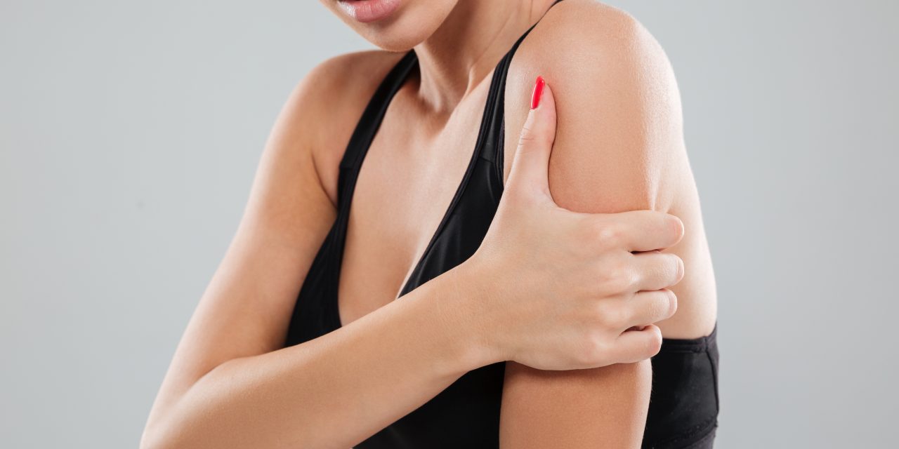
Successfully Managing SLAP Lesions
The acronym “SLAP” stands for Superior Labrum Anterior Posterior, and is used to describe a tear or detachment of the shoulder’s superior glenoid labrum; generally originating at the anchor site for the long head of the biceps tendon and extending into anterior or posterior portions of the labrum. (1) This pathology is fairly common; in fact, up to 1/4 of patients undergoing shoulder arthroscopy for any diagnosis will demonstrate a SLAP lesion. (2,3)
The glenoid labrum is a circumferential rim of fibrocartilage that surrounds the entire perimeter of the glenoid fossa. (4-6) The labrum shares some similar characteristics with the knee’s meniscus. (7) The inferior labrum is typically tightly attached to the bony glenoid rim, while the superior labrum is more “meniscus like” with a less secure union. (8) At its superior surface, the long head of the biceps tendon converges with the labrum. This provides an anchor for approximately 50% of the fibers that form the long head of the biceps tendon; the remaining fibers originate from the supraglenoid tubercle of the humerus and surrounding structures. (9)
Advertisement

The labrum serves as an attachment for tendons and ligaments and also deepens the shallow glenoid fossa by 5-9 mm. (4,10) Fifty percent of the glenoid depth is attributable to the labrum. (11) Like the meniscus of the knee, the labrum enjoys a relatively robust blood supply at birth that diminishes to sparse peripheral vascularization with advanced age. (12,13)
Labral tears may occur abruptly from injury or develop more slowly from repetitive micro trauma. (14) The forces associated with a labral injury typically include either superior compression or sudden inferior traction. (14) Traumatic onsets, including a fall or direct blow to the shoulder, are responsible for almost 1/3 of all SLAP lesions. (14) The most common mechanism of acute injury involves a fall onto an outstretched arm with the shoulder abducted and flexed forward. (14)
SLAP lesions are common in athletic populations, particularly those requiring overhead motions that encourage the biceps to “pull” the labrum from its underlying bony attachment. (1,15) Incidentally, the long head of the biceps has a dichotomous relationship with the labrum. In healthy shoulders, the long head of the biceps stabilizes the shoulder by generating compressive forces that limit translation, thereby, protecting the shoulder from anterior subluxation. (16) The biceps also depresses the humeral head to protect the labrum and subacromial contents during arm elevation. Conversely, repetitive contraction of the biceps may trigger avulsion of its labral anchor – becoming progressively more problematic as the tear progresses. (15)
Chronic SLAP lesions do not typically occur in the absence of concurrent shoulder pathology; in fact, only 28% of SLAP tears are isolated problems. (17) Chronic etiologies are often associated with rotator cuff dysfunction. Weakened and irritated rotator cuff tendons lose their ability to depress the humeral head within the glenoid cavity during arm elevation. Loss of this protective mechanism allows unchecked superior migration of the humeral head; and over time has the ability to “lift” the labrum from its attachment. (14) Not surprisingly, a high percentage (29-45%) of patients with SLAP lesions demonstrate concomitant rotator cuff pathology. (17-19) Other familiar co-morbidities include Bankart lesions (22% of SLAP cases), A/C arthrosis, instability, and supraglenoid ganglion cysts. (17,19)
SLAP lesions commonly occur in young active males. (20) Throwers may be particularly vulnerable as this activity subjects the bicep tendon and its labral attachment to significant strain. (15,21) A history of trauma or instability increases the likelihood of SLAP lesion. (3,21) However, in many cases, patients present without a history of trauma or predisposing activity. (21)
SLAP lesion complaints can vary from asymptomatic to disabling. Symptomatic patients often describe a deep, vague, non-specific shoulder pain that is provoked by overhead and cross-body activity. (20,21) Weakness and stiffness often accompany the disorder. Discomfort may limit athletic performance, particularly in overhead athletes who may complain of a “dead arm.” (20,21) Complaints of popping, clicking, grinding or catching are common. (20,21) Patients with more advanced lesions are likely to report symptoms associated with instability; i.e. (pinching, slipping, apprehension or “looseness”- especially during overhead activity)
Given that SLAP lesions are typically accompanied by additional pathology, clinical diagnosis remains challenging. (1,14,17,26) Evaluation may elicit palpatory tenderness at the origin of the long head of the biceps tendon. Range-of-motion deficits are possible, particularly in cross-body adduction and forward flexion. Motion palpation may demonstrate increased translation in patients with more advanced lesions.
No single orthopedic maneuver has been shown to reliably predict a SLAP tear. (27-29) Nevertheless, the literature is replete with no less than two dozen tests designed to help establish a diagnosis. (26,30-33)
The Biceps Load II Test has shown relatively high sensitivity and specificity in identifying isolated SLAP lesions. (26,34) The test is performed with the patient’s shoulder abducted to 120 degrees and externally rotated. The clinician stabilizes the patient’s arm while passively externally rotating until end range or patient apprehension. The patient then attempts to flex their elbow against the clinician’s resistance. An increase in pain suggests a SLAP lesion, while a decrease in apprehension or pain makes a SLAP lesion unlikely.
The Active Compression Test (O’Brien Test, Flexion-Adduction Test) may be helpful for diagnosing SLAP lesions. (35) The test is performed by having the patient position their shoulder at 90 degrees of forward elevation and 20 degrees of horizontal adduction. The patient is then asked to pronate and supinate their forearm while the examiner applies a downward force. Labral involvement is suggested when pain is reproduced while the forearm is pronated and relieved upon supination. Pain over the AC joint during this maneuver implies AC pathology and is not specific for a SLAP lesion. (36)
The Crank Test has shown utility for identifying labral pathology. (37) The test begins by having the seated or supine patient elevate their arm to 160 degrees in a scapular plane. The clinician stabilizes the shoulder with one hand and grasps the patient’s flexed elbow with the other. The clinician compresses the patient’s elbow to apply an axial load to the shoulder while performing passive internal and external rotation. Pain or “catching” suggests glenoid labrum involvement.
The Compression Rotation Test is a very similar maneuver that attempts to induce joint crepitus. This test is performed on a supine patient with their shoulder in 90 degrees abduction and 90 degrees elbow flexion while the examiner grasps the elbow and applies a compressive force into the glenohumeral joint as the shoulder is rotated internally and externally in an attempt to trap the labrum within the joint. The presence of an uncomfortable “clunk” may suggest labral tear. (38)
The Resisted Supination External Rotation Test is performed on a supine patient with their shoulder at 90 degrees of abduction, 70 degrees of elbow flexion, forearm neutral. The examiner simultaneously resists forearm supination while passively externally rotating the patient’s shoulder in an attempt to “peel back” the labrum. (36,40).
Follow this link to see a video regarding these tests and treatment of SLAP lesions
Clinicians assessing for the presence of a SLAP lesion must maintain a high degree of suspicion for co-existent pathology. Potential differential diagnostic considerations include A/C joint degeneration, strain or pathology, biceps tendinopathy, cervical radiculopathy, brachial plexus injury, fracture, Bankart lesion, dislocation, glenohumeral degeneration, instability, and most commonly, rotator cuff pathology.
Because the clinical assessment of SLAP lesion is so ambiguous, imaging is the mainstay of diagnosis. (41) Routine radiographs cannot identify the presence of a SLAP lesion; however, these studies may be useful to rule out differential considerations, including degeneration, dislocation, fracture, or pathology. Diagnostic ultrasound is a useful modality for imaging SLAP tears with an added benefit of receiving patient feedback while evaluating their injury. MRI is typically included in the initial workup for patients with suspected labral pathology. (42) The incorporation of gadolinium-enhanced MR arthrography provides the highest sensitivity (82-96%) and specificity (91-98%) for detecting SLAP lesions. (42-47)
Although MR arthrography provides the best identification of labral pathology, the accurate classification of SLAP lesions via advanced imaging is less reliable. (42) Moreover, the significance of a confirmed labral tear is debatable since this finding is present in more than half of asymptomatic, middle-aged patients. (48)
Conservative management of SLAP lesions is frequently unsuccessful. (49-51) However, the presence of a SLAP lesion does not automatically necessitate surgical intervention. Most clinicians view this structural abnormality with the same skepticism as “disc lesion”, “meniscus tear” or “rotator cuff tear”- wherein the imaged defect is not necessarily the primary contributor to the patient’s complaint. Most experts, including the American Academy of Orthopedic Surgeons, recommends a 6-12 week course of conservative management prior to considering surgical intervention. (50-52) Conservative treatment goals include pain reduction, enhancing mobility, and restoration of strength. (51)
Initial management includes anti-inflammatory medications/ modalities and cessation of provocative activities (i.e. throwing). (51) Flexibility programs are incorporated as symptoms allow. Throwing athletes are particularly predisposed to loss of glenohumeral internal rotation, and clinicians must emphasize restoration of this motion, i.e. sleeper stretch and cross-body adduction stretches. (51) Progressive strengthening of the scapular and rotator cuff musculature is implemented as tolerated. Strengthening exercises should focus on re-balancing strength between anterior (pec, upper traps) and posterior muscle groups (lower traps, serratus anterior, rhomboid). Rehabilitation must restore serratus anterior strength and proper scapular function- thoroughly addressing regional biomechanical deficits including scapular dyskinesis and upper crossed syndrome, as well as more distant sites of dysfunction including the hip and trunk. Myofascial release may be directed at the subscapularis, infraspinatus, anterior shoulder musculature and the posterior capsule. (53) Manipulation may be helpful to restore mobility to restricted cervical or thoracic segments.
References
- Knesek M, Skendzel JG, Dines JS, et al. Diagnosis and management of superior labral anterior posterior tears in throwing athletes. Am J Sports Med 2013;41(2): 444–60.
- Kim TK, Queale WS, Cosgarea AJ, McFarland EG. Clinical features of the different types of SLAP lesions: an analysis of one hundred and thirty-nine cases. J Bone Joint Surg Am. 2003 Jan. 85-A(1):66-71.
- Kampa RJ, Clasper J. Incidence of SLAP lesions in a military population. J R Army Med Corps. 2005 Sep. 151(3):171-5.
- Snyder SJ, Karzel RP, Del Pizzo W, Ferkel RD, Friedman MJ. SLAP lesions of the shoulder. Arthroscopy. 1990. 6(4):274-9.
- Moseley H, Overgaard B. The anterior capsular mechanism in recurrent anterior dislocation of the shoulder. Morphological and clinical studies with special reference to the glenoid labrum and glenohumeral ligaments. J Bone Joint Surg Br. 1962. 44B:913-27.
- Detrisac DA, Johnson LL. Arthroscopic Shoulder Anatomy: Pathological and Surgical Implications. Thorofare, NJ: Slack Inc; 1986.
- Soslowsky LJ, Flatow EL, Bigliani LU, et al. Quantitation of in situ contact areas at the glenohumeral joint: a biomechanical study. J Orthop Res 1992;10(4):524–34
- Williams RJ. Superior Labrum Tears. Medscape. http://emedicine.medscape.com/article/92512-overview#a5. Accessed Feb 12, 2016.
- Vangsness CT Jr, Jorgenson SS, Watson T, Johnson DL. The origin of the long head of the biceps from the scapula and glenoid labrum. An anatomical study of 100 shoulders. J Bone Joint Surg Br. 1994 Nov. 76(6):951-4.
- Rockwood CA, Matsen FA, Wirth MA. The shoulder, vol. 1. Philadelphia: WB Saunders; 2009.
- Howell SM, Galinat BJ. The glenoid-labral socket. A constrained articular surface. Clin Orthop Relat Res 1989;(243):122–5.
- Williams RJ. Superior Labrum Tears. Medscape. http://emedicine.medscape.com/article/92512-overview#a5. Accessed Feb 12, 2016.
- Prodromos CC, Ferry JA, Schiller AL, Zarins B. Histological studies of the glenoid labrum from fetal life to old age. J Bone Joint Surg Am. 1990 Oct. 72(9):1344-8.
- Dessaur WA. (2008). Diagnostic Accuracy of Clinical Testing for Superior Labral Anterior Posterior Lesions: A Systematic Review Journal of Orthopaedic and Sports
- Andrews JR, Carson WG Jr, McLeod WD. Glenoid labrum tears related to the long head of the biceps. Am J Sports Med. 1985 Sep-Oct. 13(5):337-41.
- Pagnani MJ, Deng XH, Warren RF, Torzilli PA, Altchek DW. Effect of lesions of the superior portion of the glenoid labrum on glenohumeral translation. J Bone Joint Surg Am. 1995 Jul. 77(7):1003-10.
- Snyder SJ, Banas MP, Karzel RP: An analysis of 140 injuries to the superior glenoid labrum. J Shoulder Elbow Surg 4: 243-248, 1995.
- Andrews JR, Carson WG. The arthroscopic treatment of glenoid labrum tears in the throwing athlete. Orthop Trans. 1984;8:44.
- Mileski RA, Snyder SJ. Superior labral lesions in the shoulder: pathoanatomy and surgical management. J Am Acad Orthop Surg. 1998;6:121–131
- David A. Cortese and Stephen J. Snyder Superior Labral Anterior-Posterior Lesions of the Shoulder THE ATHLETE’S SHOULDER (Second Edition), 2009, Pages 269-282
- Bedi A, Allen AA. Superior labral lesions anterior to posterior-evaluation and
- arthroscopic management. Clin Sports Med 2008;27(4):607–30.
- Gabel CP, Yelland M, Melloh M, et al. A modified QuickDASH-9 provides a valid outcome instrument for upper limb function. BMC Musculoskelet Disord 2009;10:161.
- Roy JS, MacDermid JC, Woodhouse LJ. Measuring shoulder function: a systematic review of four questionnaires. Arthritis Rheum 2009;61(5):623–32.
- Godfrey J, Hamman R, Lowenstein S, et al. Reliability, validity, and responsiveness of the simple shoulder test: psychometric properties by age and injury type. J Shoulder Elbow Surg 2007;16(3):260–7.
- MacDermid JC, Solomon P, Prkachin K. The shoulder pain and disability index demonstrates factor, construct and longitudinal validity. BMC Musculoskelet Dis- ord 2006;7:12.
- Cook C et al. Diagnostic accuracy of five orthopedic clinical tests for diagnosis of superior labrum anterior posterior (SLAP) lesions Journal of Shoulder and Elbow Surgery. Vol 21, Issue 1 January 2012, Pages 13–22
- Guanche CA, Jones DC: Clinical testing for tears of the glenoid labrum. Arthroscopy 19:517-523, 2003.
- Holtby R, Razmjou B: Accuracy of the Speed’s and Yergason’s tests in detecting biceps pathology and SLAP lesions: Comparison with arthroscopic findings. Arthroscopy. 20:231-236, 2004.
- McFarland EG, Kim TK, Savino RM: Clinical assessment of three common tests for superior labral anterior-posterior lesions. Am J Sports Med 30:810-815, 2002.
- Dutton Mark. Orthopaedic examination, evaluation, and intervention. New York: McGraw Hill; 2004.
- Dessaur WA. (2008). Diagnostic Accuracy of Clinical Testing for Superior Labral Anterior Posterior Lesions: A Systematic Review Journal of Orthopaedic and Sports
- Stetson WB, Templin K. The crank test, the O’Brien test, and routine magnetic resonance imaging scans in the diagnosis of labral tears. Am J Sports Med. 2002;30:806–809.
- Yang-Soo Kim, Jung-Man Ha, Kee Yong, et al. The passive compression test: A new clinical test for superior labral tears of the shoulder. Am J Sports Med, 2007;35(9):1489-94.
- Kim SH et al. Biceps Load Test II: A Clinical test for SLAP lesions of the Shoulder. Arthroscopy 2001
- O’Brien SJ, Pagnani MJ, Fealy S, McGlynn SR, Wilson JB. The active compression test: a new and effective test for diagnosing labral tears and acromioclavicular joint abnormality. Am J Sports Med. 1998;26:610–613
- Dodson CC, Altchek DW. SLAP Lesions: An Update on Recognition and Treatment. JOSPT February 2009, Volume 39 Number 2
- Dessaur WA. (2008). Diagnostic Accuracy of Clinical Testing for Superior Labral Anterior Posterior Lesions: A Systematic Review Journal of Orthopaedic and Sports
- Snyder SJ, Karzel RR, Del Pizzo W, Ferkel RD, Friedman MJ. SLAP lesions of the shoulder. Arthroscopy. 1990;6:274–279
- Myers TH, Zemanovic JR, Andrews JR. The resisted supination external rotation test: a new test for the diagnosis of superior labral anterior posterior lesions. Am J Sports Med. 2005;33:1315–1320.
- George D. Chloros et al., Imaging of Glenoid Labrum Lesions Clin Sports Med 32 (2013) 361–390
- Williams RJ. Superior Labrum Tears. Medscape. http://emedicine.medscape.com/article/92512-overview#a5. Accessed Feb 12, 2016.
- Fallahi F, Green N, Gadde S, Jeavons L, Armstrong P, Jonker L. Indirect magnetic resonance arthrography of the shoulder; a reliable diagnostic tool for investigation of suspected labral pathology. Skeletal Radiol. 2013 Sep. 42(9):1225-33.
- Modi CS, Karthikeyan S, Marks A, Saithna A, Smith CD, Rai SB, et al. Accuracy of abduction-external rotation MRA versus standard MRA in the diagnosis of intra-articular shoulder pathology. Orthopedics. 2013 Mar. 36(3):e337-42.
- Magee T, Williams D, Mani N. Shoulder MR arthrography: which patient group benefits most? AJR Am J Roentgenol 2004;183(4):969–74.
- Monu JU, Pope TL Jr, Chabon SJ, Vanarthos WJ. MR diagnosis of superior labral anterior posterior (SLAP) injuries of the glenoid labrum: value of routine imaging without intraarticular injection of contrast material. AJR Am J Roentgenol. 1994 Dec. 163(6):1425-9.
- Waldt S, Burkart A, Lange P, et al. Diagnostic performance of MR arthrography in the assessment of superior labral anteroposterior lesions of the shoulder. AJR Am J Roentgenol. 2004 May. 182(5):1271-8.
- Schwartzberg R, Reuss BL, Burkhart BG et al. High Prevalence of Superior Labral Tears Diagnosed by MRI in Middle-Aged Patients With Asymptomatic Shoulders. Journal of Sports Medicine. January 2016 vol. 4 no. 1
- Williams RJ. Superior Labrum Tears. Medscape. http://emedicine.medscape.com/article/92512-treatment. Accessed Feb 12, 2016
- Mileski RA, Snyder SJ. Superior labral lesions in the shoulder: pathoanatomy and surgical management. J Am Acad Orthop Surg. 1998 Mar-Apr. 6(2):121-31
- Dodson CC, Altchek DW. SLAP Lesions: An Update on Recognition and Treatment. JOSPT February 2009, Volume 39 Number 2
- Franceschi F, Longo UG, Ruzzini L, et al. No advantages in repairing a type II su- perior labrum anterior and posterior (SLAP) lesion when associated with rotator cuff repair in patients over age 50: a randomized controlled trial. Am J Sports Med 2008;36(2):247–53.
- Hammer W. Conservative Treatment of SLAP Tears. Dynamic Chiropractic. September 9, 2010, Vol. 28, Issue 19


