


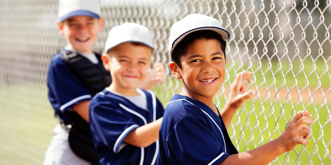
Little League Elbow
The term, “Little League Elbow” (LLE) was first coined in 1960. (1) The diagnosis is now used to describe a predictable progression of overuse injuries that affect the medial elbow of children and adolescents. (2) The primary culprit for LLE is repetitive valgus overload, causing painful osteocartilaginous injury or ligamentous irritation. (3)
Medial Elbow Structure
The medial elbow is statically stabilized by its bony architecture, joint capsule, and medial (ulnar) collateral ligament (MCL). (4) The MCL is a ligamentous complex consisting of three bundles (anterior oblique, posterior oblique, and transverse) that bind the ulna to the humerus and provide the elbow’s primary static support against valgus stress. (4)
Advertisement

The MCL and medial elbow receive dynamic support from the flexor-pronator muscles that originate on the medial epicondyle. The “common flexor” tendon consists of the flexor carpi radialis, palmaris longus, flexor digitorum superficialis, and flexor carpi ulnaris. (4-7) The pronator teres originates on the supracondylar ridge of the medial epicondyle, slightly above the common flexor tendon, with a secondary origin on the ulna coronoid process. (7)
A brief osseous terminology refresher may be helpful before discussing developmental pathology:
- Physis – the cartilaginous growth plate (secondary ossification center) near the end of a long bone.
- Epiphysis – the “cap” of bone located between the growth plate (physis) and joint cartilage.
- Apophysis – an outgrowth from the host bone that serves as an attachment for tendons or ligaments. The apophysis is attached to the host bone by a growth plate.
A “traction apophysitis,” like little league elbow, affects growth plates, which, in children, are believed to be 2-5 times weaker than the surrounding tissue. (8,11) The elbow has six growth plates that typically appear and fuse at different ages (9):
- capitellum (appears 1-2 years old, fuses at 10-15)
- radial head (appears 2-5 years old, fuses at 12-16)
- trochlea (appears 8-10 years old, fuses at 10-18)
- olecranon (appears 8-10 years old, fuses at 13-16)
- lateral epicondyle (appears 8-13 years old, fuses at 12-16)
- medial epicondyle (appears 2-8 years old, fuses at 15-17)
In evaluating the growth centers about the elbow among individuals, substantial variability exists, but it is important to note that there is considerably less variability in each individual from side to side. The medial epicondyle is predictably the one of last ossification center to close, beginning the process at age seven, with complete fusion around age 15 in boys and 13 in girls. (9,10)
Medial column injury is the most common overuse elbow malady in young athletes, particularly throwers. (11) The act of overhand pitching places compressive stress on the lateral compartment of the elbow, and tremendous valgus stress on the medial elbow. (12-14) This causes tensile distraction on the medial column structures, including the medial epicondyle, medial ulnar crest, and MCL. (13) These forces are most intense during the late cocking and early acceleration phases of throwing. (4,5) Stress peaks during late cocking when the elbow is held near 90 degrees. (15) Valgus torque forces are appreciable in adolescents (18-28 Nm), and professionals must endure up to five times more stress. (90-120 Nm). (15) For crude illustration, professional pitching equates to placing your upper arm in a vice, medial side up, then dropping 20-25 lbs. on your forearm, ten centimeters down from the elbow, and repeating 100 plus repetitions every fifth day.
Medial elbow pain affects 20-58% of youth baseball players, with increasing incidence throughout adolescence and adulthood. (16-18). Injuries are somewhat predictable based on the patient’s bony development. (13)
- In skeletally immature patients, the growth plate at the base of the medial epicondyle is the weakest link in the medial column. (19,20) Not surprisingly, stress in skeletally immature patients leads to traction apophysitis of the medial epicondyle. (13)
- As the medial apophysis growth plate fuses to the humerus, vulnerability shifts to the bone-ligament interface, so traction avulsion injuries become more likely. (20)
- Following skeletal maturation, susceptibility transitions from bone to soft tissues, where injuries to the MCL prevail. (13,20)
LLE is most common in baseball pitchers but also affects other position players. Elbow overuse injuries often strike stronger (more skilled) players, who are habitually chosen as pitchers and given abundant playing time. (21) To compound the issue, these better players are often channeled into single sport participation with year-round training and longer competitive seasons.
Pitch count is the primary determinant for youth medial elbow damage. (22) Children throwing over 600 pitches per season more than double their risk for elbow injury. (23) Historically, parents and coaches have been educated that curveballs and other forms of off-speed pitches should be avoided until high school. More contemporary research suggests that other variables, such as pitch count, rest between pitch outings, and proper body mechanics, are more important considerations. Contrary to popular belief, pitch type does not affect elbow injury rates; in particular, curveballs are no more hazardous than any other pitch. (24,42)
While most cases of LLE strike baseball players, the malady frequently affects athletes from other sports, including softball, tennis, racquetball, handball, javelin, water polo, gymnastics, and weight lifting. (18)
Injury Presentation
The classic presentation for LLE is a young or adolescent baseball or softball player complaining of medial elbow pain in their throwing arm. Symptoms often present in mid to late-season, as pitch counts elevate, cumulative workload increases, and rest is diminished. Alternatively, this may be seen after a single game with increased or pitch count without appropriate rest. (22,24) Players may avoid complaining of pain, because of their desire to compete, and instead express frustration because of decreased velocity, accuracy, and distance. (24) This is often complicated by parents and coaches urging the individual to “strengthen” their arm for greater velocity and distance, while encouraging increased practice time to improve accuracy.
Clinical evaluation will universally demonstrate point tenderness over the medial epicondyle. (25) This is often associated with localized swelling, elbow stiffness and, sometimes, the inability to achieve full extension. Resisted wrist flexion and pronation will likely exacerbate symptoms. (25) The Golfer’s elbow test is predictably provocative. In more advanced cases, loss of full elbow extension is possible. (25) Flexion contractures in children and adolescents suggest significant damage. (26)
Clinicians should assess valgus stability with the moving valgus stress test. This test is performed by passively flexing and extending the patient’s elbow while applying a valgus force through the joint line. LLE patients may report apprehension or discomfort; however, instability or giving way at mid-range suggests medial ulnar collateral ligament laxity. (27)
The Milking test is another useful maneuver to help identify MCL laxity or disruption. The test is performed by having the seated patient elevate and externally rotate their shoulder, as though they were asking a question. With their elbow flexed at least 90 degrees, the patient supinates their forearm, and extends their thumb so that it is pointing directly backward. The clinician stands behind the patient and repeatedly pulls (“milks”) the patient’s thumb while the clinician’s other hand stabilizes the elbow and palpates the medial joint line. This movement places valgus stress on the elbow. Reproduction of pain, apprehension, or laxity suggests MCL involvement, particularly of the anterior bundle. (28)
Evaluation
Clinicians should perform a neurologic evaluation of the upper extremity with emphasis on the ulnar nerve. Cubital tunnel tenderness or a positive Tinel sign suggests ulnar neuropathy.
Global biomechanical deficits can place undue stress on peripheral joints. The medial elbow is a frequent casualty of core and hip problems. The throwing arm can be likened to a catapult connected to a base (shoulder) that is securely mounted to a foundation (core). Repeatedly firing a catapult that does not have a mechanically sound base or foundation will lead to a loss of velocity, accuracy, and integrity of the components. Likewise, prior or concurrent injuries in any other part of the kinetic chain can alter throwing mechanics and exploit other weak links in the system. For most cases of LLE, the medial epicondyle was merely the (dysfunctional) chain’s weakest biomechanical link, and first to exhibit symptoms.
Accordingly, a comprehensive clinical evaluation must screen for signs of remote dysfunction, including scapular dyskinesis, dysfunctional breathing, spinal instability, or hip abductor weakness. Please see the corresponding ChiroUp protocols for detailed descriptions of each deficit.
Radiographs may be appropriate if the patient is unresponsive to initial conservative treatment, or if alternate pathology is suspected. Patients presenting with significant swelling and fixed flexion positioning should be imaged without delay to rule out avulsion or epicondylar fracture. (25) Standard elbow radiographs include AP and images. The addition of axial and medial oblique radiographs, as well as AP valgus stress projections, may provide additional information when specifically addressing LLE. (29)
Radiographs of young elbows, without exception, demonstrate irregularity of the medial epicondylar physis. (19,30) This irregularity typically decreases sharply after age 14. (30) Changes of chronic medial column stress include widening of the medial epicondylar physis, hypertrophy of the medial epicondyle and cortical thickening of the medial aspect of the distal humerus. (29) Approximately 10% of throwers will demonstrate some degree of medial epicondyle fragmentation on plain films. (30) Often bilateral radiographs are necessary to evaluate for asymmetry of the osseous structures and adjacent growth plate for subtle differences. Radiographic findings of acute injury include fracture and physeal widening.
Advanced imaging can help confirm diagnostic suspicions and potentially identify occult osteocartilaginous pathology, ligament injury and associated myotendinous findings. (19) Ultrasound and MRI frequently demonstrate findings characteristic of LLE, including medial epicondylar hypertrophy (90-100%), physeal separation (50-70%), and osseous fragmentation (15-40%). (30,31) Patients with characteristic imaging lesions have a significantly greater likelihood for elbow pain (30); however, those imaging findings may not necessarily change the prescribed clinical management and outcomes. (31)
Differential Diagnoses
The differential diagnosis for LLE includes dislocation, fracture, avulsion, infection, neoplasm, exostosis, osteochondritis dissecans, meniscoid lesion, cervical radiculopathy (C8), ulnar neuropathy, aka cubital tunnel syndrome, posteromedial impingement, and sprain/strain, particularly of the flexor/pronator tendon, medial ulnar collateral ligament, or joint capsule.
LLE typically responds well to conservative management. (4,5,32) Rest and anti-inflammatory modalities are the mainstays of early treatment. Similar to other overuse injuries, elimination of the inciting event is the key to management. (33) Although unpopular, the most expedient management requires that the athlete cease all throwing activity. (5,32,34) LLE patients may need to selectively restrict throwing for up to four to six weeks. (21) Athletes should otherwise remain active. Splinting or wrist braces may help limit elbow strain in acute or refractory cases. (34)
Treatment
Initial rehab includes range of motion exercises accompanied by anti-inflammatory measures. (5) Patients may ice their medial elbow for ten minutes, four times per day. Counterirritant creams may provide palliative relief. Neurodynamic techniques for the ulnar and median nerves may be beneficial.
Early strengthening could include extension and supination-based exercises until the patient can tolerate flexion and pronation. (35) Eventually, rehab should progress to include isotonic strengthening of the wrist flexors, extensors, pronators, and supinators with lower resistance/high repetition exercises. (35) Eccentric strengthening exercises may be appropriate, particularly when there is tendon involvement in more mature LLE patients. (43) The “Reverse Tyler Twist” exercise, utilizing a Theraband Flexbar is a novel approach to eccentric strengthening of the wrist flexors. (44,45)
Long-term rehab should incorporate strengthening of the flexor carpi ulnaris and flexor digitorum superficialis, both of which provide secondary support to the MCL. This fortification is relevant because the same forces that caused LLE will inevitably lead to progressive stretching, laxity, and disruption of the MCL.
Any efforts to repair and strengthen the elbow will likely fail without optimizing function of the more proximal shoulder girdle and core. Scapular stabilization exercises should target the lower trapezius and serratus anterior. (36) Shoulder external rotation and retraction exercises have been shown to help rehabilitate and prevent elbow injuries. (37) Specific exercises could include a Brugger with band, low row, and lawnmower. (36,38)
Conclusion
Medial elbow injuries can be triggered or exacerbated when the athlete fails to properly engage the core and leg muscles responsible for balance and power generation. (39) Appropriate core and hip stability exercises include bridging, posterior lunges, semi-stiff deadlift, and clam progressions. Single leg proprioceptive exercises may be beneficial. In addition, a thorough assessment of shoulder, trunk and lower extremity flexibility, and providing pertinent stretches to address any deficiencies, is indicated.
A gradual return to throwing may be initiated when there is no longer point tenderness over the medial elbow. (32) Patients should not advance training more than 10% per workout and may return to play when they reach sport-specific levels of performance. (32)
Following return to activity, monitoring pitch count and rest days is crucial. The USA Baseball Medical and Safety Advisory Committee has published widely endorsed age-specific recommendations for pitch limits per game, week, season, and year. View the Pitch Smart® guidelines here. (40)
Coaches and parents must understand the importance of limiting activity and should follow guidelines for the player’s skeletal age as opposed to league age, since often, the most skilled (and most vulnerable) players “play up.” (41) It is quite common for players to have individual pitching coaches, and, at the high school level, play on a competitive/travel team, in addition to a league high school program. It is imperative that communication exists between all coaches and the parents of the player to ensure that the physical health of the youth athlete is the top priority.
Young pitchers should not pitch on consecutive days or in multiple games per day. Pitchers should not be utilized as catchers on “off days.” Players should avoid pitching on multiple teams with overlapping seasons. Players should have a 2-4 month “off-season” that does not involve throwing. Young players should stay away from radar guns or other measurement devices that encourage them to consistently throw harder.
Oral analgesia, ice, and modalities may be used as early anti-inflammatory agents. (5,34,35) However, patients should understand that some degree of inflammation is necessary for healing, and over-the-counter pain medication must never be employed solely to enable continuation of the offending activity. (4) Steroid injections for the management of LLE should be avoided, as they may create further ligament or cartilage damage. (4)
“LLE Treatment is conservative, and there is little to no role for operative intervention.” (32) However, orthopedic referral is warranted for patients who fail a trial of conservative care, demonstrate evidence of MCL disruption, or have greater than 2 mm of medial epicondyle separation/avulsion. (21) Stable avulsion fractures with 2-5 mm of separation are generally immobilized with a long-arm cast, while those greater than 5 mm may require surgical intervention. (33,34)
References
1. Brogdon BG, Crow NE. Little leaguer’s elbow. Am J Roentgenol Radium Ther Nucl Med 1960; 83:671–5.
2. Kiery A. Braithwaite, Kelley W. Marshall. The Skeletally Immature and Newly Mature Throwing Athlete. Radiol Clin N Am 54 (2016) 841–855.
3. Frush TJ, Lindenfeld TN. Peri-epiphyseal and over-use injuries in adolescent athletes. Sports Health 2009;1(3):201–11.
4. Gregory B, Nyland J. Medial elbow injury in young throwing athletes. Muscles Ligaments Tendons J. 2013 Jul 9;3(2):91-100. doi: 10.11138/mltj/2013.3.2.91. PMID: 23888291; PMCID: PMC3711707.
5. Park MC, Ahmad CS. Dynamic contributions of the flexor-pronator mass to elbow valgus stability. J Bone Joint Surg Am 2004;86-A(10):2268–74.
6. Stoller DW, editor. Magnetic resonance imaging in orthopaedics and sports medicine. Lippincott Williams & Wilkins; 2007. Link
7. Ciccotti MC, Schwartz MA, Ciccotti MG. Diagnosis and treatment of medial epicondylitis of the elbow. Clinics in sports medicine. 2004 Oct 1;23(4):693-705. Link
8. Schwab GH, Bennett JB, Woods GW, Tullos HS. Biomechanics of elbow instability: the role of the medial collateral ligament. Clin Orthop Relat Res. 1980;(146):42–52.
9. Klingele KE, Kocher MS. Little league elbow: valgus overload injury in the paediatric athlete. Sports Med 2002;32(15):1005–15.
10. Patel B, Reed M, Patel S. Gender-specific pattern differences of the ossification centers in the pediatric elbow. Pediatr Radiol 2009;39(3):226–31.
11. Jaimes C, Chauvin NA, Delgado J, et al. MR imaging of normal epiphyseal development and common epiphyseal disorders. Radiographics 2014;34(2): 449–71.
12. Dugas JR. Valgus extension overload: diagnosis and treatment. Clin Sports Med 2010;29(4):645–54.
13. Wei AS, Khana S, Limpisvasti O, et al. Clinical and magnetic resonance imaging findings associated with Little League elbow. J Pediatr Orthop 2010; 30(7):715–9.
14. Vamsi K. Kancherla, Nicholas M. Caggiano, Kristofer S. Matullo, Elbow Injuries in the Throwing Athlete. Orthop Clin N Am 45 (2014) 571–585
15. Sabick MB, Torry MR, Lawton RL, Hawkins RJ. Valgus torque in youth baseball pitchers: A biomechanical study. J Shoulder Elbow Surg. 2004;13(3):349–355
16. Lyman S, Fleisig GS, Waterbor JW, et al. Longitudinal study of elbow and shoulder pain in youth baseball pitchers. Med Sci Sports Exerc 2001;33(11): 1803–10.
17. Lyman S, Fleisig GS, Andrews JR, et al. Effect of pitch type, pitch count, and pitching mechanics on risk of elbow and shoulder pain in youth base- ball pitchers. Am J Sports Med 2002;30(4):463–8.
18. Chen AL, Youm T, Ong BC, Rafii M, Rokito AS. Imaging of the elbow in the overhead throwing athlete. Am J Sports Med. 2003;31(3):466–473.
19. Kijowski R, Tuite MJ. Pediatric throwing injuries of the elbow. Semin Musculoskelet Radiol 2010;14(4):419–29.
20. Kijowski R, Tuite MJ. Pediatric throwing injuries of the elbow. Semin Musculoskelet Radiol 2010;14(4):419–29.
21. Little League Baseball—The Learning Curve. http://www.littleleague.org/Assets/ forms_pubs/media/UNCStudy.pdf.
22. Lyman S, Fleisig GS, Andrews JR, et al. Effect of pitch type, pitch count, and pitching mechanics on risk of elbow and shoulder pain in youth base- ball pitchers. Am J Sports Med 2002;30(4):463–8.
23. Lyman S, Fleisig GS, Waterbor JW, et al. Longitudinal study of elbow and shoulder pain in youth baseball pitchers. Med Sci Sports Exerc 2001;33(11): 1803–10
24. Kobayashi K, Burton KJ, Rodner C, et al. Lateral compression injuries in the pediatric elbow: Pan- ner’s disease and osteochondritis dissecans of the capitellum. J Am Acad Orthop Surg 2004;12(4): 246–54.
25. Vamsi K. Kancherla, Nicholas M. Caggiano, Kristofer S. Matullo, Elbow Injuries in the Throwing Athlete. Orthop Clin N Am 45 (2014) 571–585
26. Todd S. Ellenbecker, Michael Reinold, Cory O. Nelson. Clinical Concepts for Treatment of the Elbow in the Adolescent Overhead Athlete. Clin Sports Med 29 (2010) 705–724
27. O’Driscoll SW, Lawton RL, Smith AM. The “moving valgus stress test” for medial collateral ligament tears of the elbow. Am J Sports Med 2005;33(2): 231–9.
28. Hariri S, Safran MR. Ulnar collateral ligament injury in the overhead athlete. Clin Sports Med. 2010;29(4):619–644
29. Vamsi K. Kancherla, Nicholas M. Caggiano, Kristofer S. Matullo, Elbow Injuries in the Throwing Athlete. Orthop Clin N Am 45 (2014) 571–585
30. Otoshi, K., Kikuchi, S., Kato, K., Sato, R., Igari, T., Kaga, T., & Konno, S. (2017). Age-Specific Prevalence and Clinical Characteristics of Humeral Medial Epicondyle Apophysitis and Osteochondritis Dissecans: Ultrasonographic Assessment of 4249 Players. Orthopaedic Journal of Sports Medicine
31. Hang DW, Chao CM, Hang Y-S. A clinical and roentgenographic study of Little League elbow. Am J Sports Med 2004;32(1):79–84.
32. Andelman S, DiPrinzio E, Hausman M. Elbow injuries in the pediatric athlete. Annals of Joint. 2018 Mar 23;3(3). Link
33. Osbahr DC, Chalmers PN, Frank JS, et al. Acute, avulsion fractures of the medial epicondyle while throwing in youth baseball players: a variant of Lit- tle League elbow. J Shoulder Elbow Surg 2010; 19(7):951–7.
34. Lawrence JT, Patel NM, Macknin J, et al. Return to competitive sports after medial epicondyle frac- tures in adolescent athletes: results of operative and nonoperative treatment. Am J Sports Med 2013;41(5):1152–7.
35. Todd S. Ellenbecker, Michael Reinold, Cory O. Nelson. Clinical Concepts for Treatment of the Elbow in the Adolescent Overhead Athlete. Clin Sports Med 29 (2010) 705–724
36. Kibler WB. The role of the scapula in athletic shoulder function. Am J Sports Med 1998;26(2):325–37.
37. McCabe RA, Orishimo KF, McHugh MP, et al. Surface electromyographic analysis of the lower trapezius muscle during exercises performed below ninety degrees of shoulder elevation in healthy subject. N Am J Sports Phys Ther 2007;2(1):34–43.
38. Kibler WB, Sciascia AD, Uhl TL, et al. Electromyographic analysis of specific exercises for scapular control in early phases of shoulder rehabilitation. Am J Sports Med 2008;36:1789–98.
39. Davis JT, Limpisvasti O, Fluhme D, et al. The effect of pitching biomechanics on the upper extremity in youth and adolescent baseball pitchers. Am J Sports Med 2009;37:1484-91.
40. PitchSmart USA. Guidelines for Youth and Adolescent Pitchers. Accessed 12/17/19 from: https://www.mlb.com/pitch-smart/pitching-guidelines
41. DeLee J, Drez D, Miller MD. DeLee & Drez’s ortho- paedic sports medicine: principles and practice. 3rd edition. Philadelphia: Saunders/Elsevier; 2010.
42. Oliver GD et al. Effects of a Simulated Game on Upper Extremity Pitching Mechanics and Muscle Activations Among Various Pitch Types in Youth Baseball Pitchers. J Pediatr Orthop. 2019 Sep;39(8):387-393.
43. 41. Jean-François Kaux, Current opinions on tendinopathy Journal of Sports Science and Medicine (2011) 10, 238-253
44. Page P. A New Exercise For Tennis Elbow That Works! N Am J Sports Phys Ther. Sep 2010; 5(3): 189–193.
45. Tyler T.F., et al. , Addition of isolated wrist extensor eccentric exercise to standard treatment for chronic lateral epicondylosis: A prospective randomized trial. J Shoulder Elbow Surg. 2010; 19(6):917–922


