


Current Best Practices for Assessing and Managing Lumbar Spine Stenosis
A new study in Archives of Physical Medicine and Rehabilitation concluded: “For patients with neurogenic claudication due to lumbar spine stenosis (LSS), a comprehensive conservative program demonstrated superior, large and sustained improvements in walking ability and can be a safe non-surgical treatment option.”(53)
Chiropractic physicians who deliver “comprehensive conservative care” are validating their merit in the new fee-for-outcomes world. The following article will focus on current best practices for assessing and managing lumbar spine stenosis.
Advertisement
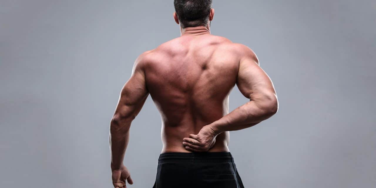
Lumbar Spine Stenosis
Lumbar spine stenosis describes the narrowing of the central spinal canal, lateral recess, neural foramina or any combination of these sites. Stenosis is subclassified by the anatomic site of narrowing, i.e. central canal stenosis, lateral recess stenosis or foraminal stenosis. Neural encroachment by the surrounding bone and soft tissue can induce subsequent “neurogenic claudication” or radiculopathy with resulting leg pain, numbness, paresthesia, weakness, and difficulty walking.
Spinal stenosis may be classified as either primary, caused by congenital/ developmental abnormalities, or secondary/acquired, arising from degenerative changes, trauma, infection or surgery. (1) The process of degenerative stenosis results from chronic accumulation of mechanical stresses, causing intervertebral disc degeneration, facet hypertrophy and ligamentum flavum fibrotic hypertrophy, or “wrinkling” (i.e., as discs thin, the ligamentum flavum “wrinkles” in much the same way that a pant leg would wrinkle if the leg shortened). (2,49) Central spinal stenosis most typically affects the three lowest lumbar levels, and foraminal stenosis most commonly involves the L5 nerve root, as the L5-S1 foramen has the least free space. (2,3)
Symptoms are thought to arise from a compressive vascular compromise, or, in more severe cases, from direct pressure. (4) The available space in the central canal and lateral recess decreases in extension (standing) and increases with flexion or axial distraction. (5) The intervertebral foramina undergo a 15% decrease surface area in extension and at 12% increase surface area in flexion. (6) Activities that require spinal extension or increase the metabolic demands of the neurologic tissues are more likely to generate symptoms, i.e., standing and walking. (7)
Reduction before Impingement
Cadaveric studies suggest that the lumbar nerve roots can tolerate up to a 70% reduction in IVF diameter before undergoing impingement. (47) The critical dimensions for lumbar neuroforaminal encroachment have been identified as posterior disc thinning to less than 4 mm of height or reduction of the IVF height to less than 15mm. (48)
The reported incidence of spinal stenosis in patients with back pain is 3-14%, but not all patients meeting the clinical definition of stenosis will report symptoms. (10) Patients with anatomic lumbar spine stenosis may fall somewhat unpredictably on a spectrum between asymptomatic and severely disabled. No relevant association has been shown between the MRI finding of “stenosis” and the severity of the patient’s pain. (51) The course of symptomatic individuals continues on a variable course with 50-70% of spinal stenosis patients remaining stable, while the remainder are divided fairly equally into groups that inconsistently improve or worsen. (8-11)
Symptoms
Symptoms of primary stenosis (non-degenerative acquired stenosis) may present early in life, while degenerative stenosis is uncommon before age 50, rarely appearing before the sixth or seventh decade. (12) Stenosis is the most common reason for lumbar spine surgery in those over 65. (13)
Symptomatic spinal stenosis often presents as chronic lower back pain with transient neurogenic claudication symptoms involving the buttock and lower extremity. Patients may report bilateral and symmetrical pain, paresthesia, numbness, fatigue, heaviness and/or weakness in their legs. Lower extremity complaints are generally perceived as more bothersome than local symptoms. Symptoms are bilateral in almost 7 out of 10 patients. (13,45) Although many other causes of lower back pain are exacerbated by prolonged sitting, lumbar spine stenosis patients report progressively increasing symptoms from standing or walking and relief while sitting. Patients may report diminished symptoms when walking with a shopping cart or lawn mower, which induces slight lumbar flexion. Walking downhill tends to increase the patient’s lumbar lordosis and is generally more uncomfortable than walking uphill. As the condition progresses, patients often adopt a slightly forward flexed posture with knee, hip and trunk flexion, i.e. “Simian” stance. (14) Patients are generally more comfortable sleeping on their sides in a fetal position, as opposed to lying flat. (15) Progression of stenosis may result in a wide-based gait, exercise intolerance and major lifestyle restrictions. Patients with more significant cauda equina compression may report urinary incontinence. (16,46)
Stenosis involving the lateral recess or intervertebral foramina presents with symptoms that are more likely unilateral and radicular in nature. Konno, et al., offered a self-administered, self-reported history questionnaire that demonstrated 84% sensitivity and 78% specificity for the diagnosis & anatomic differentiation of lumbar stenosis. (17)
Konno Stenosis Questionnaire
Do you Have?
1. Numbness and/or pain in the thighs down to the calves and shins.
2. Numbness and/or pain increases after walking and is relieved by resting.
3. Standing for a while brings on numbness and/or pain in the thighs down to the calves and shins.
4. Numbness and/or pain are reduced by bending forward.
5. Numbness is present in both legs.
6. Numbness is present in the soles of both feet.
7. Numbness arises around the buttocks.
8. Numbness is present, but pain is absent.
9. A burning sensation arises around the buttocks.
10. Walking nearly causes urination.
(Affirmative answers to questions 1-4 predict the presence of lumbar spinal stenosis, with questions 5-10 differentiating between central and lateral stenosis. Low positive scores on questions 5-10 suggest lateral recess involvement, while higher scores on these questions suggest central canal stenosis.)
Evaluation
Clinical evaluation will generally demonstrate a global reduction in lumbar mobility, particularly in extension. (18,19) The degenerative changes of lumbar spine stenosis are often accompanied by intersegmental restriction and significant myofascial involvement. (20) Orthopedic maneuvers that require extension are intended to temporarily induce neurogenic claudication and are likely to provoke symptoms. Kemp’s test has been shown to be a reliable predictor of symptom severity. (21) Other useful maneuvers include Sphinx test (sustained prone extension), Nachlas/ prone knee flexion and Yeoman test. Straight leg raise is more likely to be positive in cases of lateral recess or foraminal stenosis. Neurologic findings may include motor weakness or sensory changes, which become more evident with provocative maneuvers. (22) Diminished or absent reflexes are common in the aging population and are present in approximately half of stenosis patients. (22)
Clinicians should be cautious to rule out vascular claudication, which is characterized by five “Ps”: pulselessness, paralysis, paresthesia, pallor, and pain. Vascular claudication patients will often report symptoms that are provoked by walking and relieved by simply standing. Vascular claudication would likely be induced when the patient rides a recumbent bicycle, whereas neurogenic patients could tolerate this activity.
Radiology
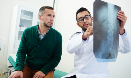
Weight-bearing radiographs are necessary to confirm the diagnosis of lumbar spine stenosis. A spinal canal diameter between 10 and 12 mm is defined as “relative stenosis,” and a diameter of less than 10 mm defines “absolute stenosis.” (23) Up to 20% of asymptomatic patients meet the radiographic criteria for the diagnosis of lumbar spine stenosis. (24,25) Radiographs frequently demonstrate associated degenerative changes. MRI may help clarify the source of neural compression but is generally reserved for those patients who have reached a stage of symptomatic or functional impairment such that the clinician is considering surgical options. (26)
The differential diagnosis of lumbar spine stenosis includes vascular claudication, peripheral neuropathy, lumbar radiculopathy, disc lesion, lumbar DDD/DJD, mechanical low back pain, myofascial pain, hip pathology, fracture, infection, neoplasm, compression fracture, hip pathology or rheumatologic disease (polymyalgia rheumatica, polymyositis, etc).
Treatment
Conservative care has been shown to help some patients with LSS. Schneider (50) identified four factors that help identify LSS patients who are most likely to respond to conservative manual therapy:
- lower initial VAS and disability scores
- relatively younger age
- radicular symptoms described as “pain”, as opposed to paresthesia or weakness
- higher body mass index (BMI)
Goals for the conservative management of lumbar spine stenosis include restoring mobility and function, decompressing neural structures and releasing perineural adhesions. (27) Rehabilitation programs should include manual therapy, stretching and endurance strengthening exercises to retard de-conditioning. (28-30) Spinal manipulation has been advocated for stenosis patients, although clinicians must be cognizant of relative contraindications from concurrent degenerative changes. (31,32) Distraction manipulation, i.e., Cox, may be a useful tool, particularly in those where HVLA manipulation is contraindicated. (33) Lumbar traction or decompression therapy shows varying degrees of long-term benefit. Nerve mobilization techniques may help to release perineural adhesions. Sciatic mobilization may be initiated by performing a straight leg raise to tolerance and then rhythmically moving the ankle through plantar flexion and dorsiflexion while gradually increasing sciatic stretch. (34) A comparative study of patients with lumbar spine stenosis found that acupuncture was significantly more effective than medication. (52)
Exercise
Exercise prescriptions should complement the treatment goals of increasing muscular, segmental and neurodynamic mobility. Exercise programs should include cat/camel and sciatic nerve flossing exercises. (35) Home stretching exercises may be necessary for hypertonic hamstring, psoas, and lumbar musculature. Recumbent cycling is usually tolerated and may help maintain aerobic function and overall fitness. Patients should limit provocative activities, including heavy lifting or those that induce extension, i.e., prolonged standing or overhead activity. Patients who are forced to stand may find relief by slightly elevating one foot on a stool, cabinet, bar rail, etc. The use of a rigid brace should be discouraged, as this will likely induce extension and provoke symptoms. (36) Inversion tables may provide palliative relief.
Medical management may include the use of NSAIDs, oral corticosteroids, gabapentin, diclofenac, and tricyclic antidepressants. (37,38) The use of epidural steroid injections is controversial, with most studies showing no or only little short-term benefit. (39-41) Patients who continue to experience debilitating symptoms and loss of function in spite of conservative efforts may be candidates for decompression by laminectomy and/ or spinal fusion surgery. (42-44)
References
1. Ciricillo SF, Weinstein PR. Lumbar spinal stenosis. West J Med. 1993 Feb;158(2):171–7.
2. Jenis LG, An HS. Spine update. Lumbar foraminal stenosis. Spine. 2000;25(3):389–94.
3. Amundsen T, Weber H, Lilleås F, Nordal HJ, Abdelnoor M, Magnaes B. Lumbar spinal stenosis. Clinical and radiologic features. Spine 1995 May 15; 20(10):1178-86.
4. Jespersen SM, Hansen ES, Høy K, Christensen KO, Lindblad BE, Ahrensberg JJ, et al. Two-level spinal stenosis in minipigs. Hemodynamic effects of exercise. Spine. 1995;20(24):2765–73.
5. Schönström N, Lindahl S, Willén J, Hansson T. Dynamic changes in the dimensions of the lumbar spinal canal: an experimental study in vitro. Journal of orthopaedic research. 1989;7(1):115–21.
6. Inufusa A, An HS, Lim TH, Hasegawa T, Haughton VM, Nowicki BH. Anatomic changes of the spinal canal and intervertebral foramen associated with flexion-extension movement. Spine. 1996 Nov 1;21(21):2412–20.
7. Schonstrom NLSWJHT. Dynamic changes in the dimensions of the lumbar spinal canal: an experimental study in vitro. J Orthop Res. 1989;7:115–121
8. Johnsson KE, Rosen I, Uden I. The natural course of lumbar spinal stenosis. Clin Orthop. 1992;279:82–86.
9. Herno A. Spinal Stenosis without deformity: Nonoperative treatment. In: Herkowitz HH, Dvorak JJ, Bell G, Nordin M, Grob DD, editors. The Lumbar Spine. Philadelphia: Lippincott Williams & Wilkins; 2004. pp. 490–4.
10. www.Spinal Stenosis.org (Retrieved 9/28/13)
11. Jenis LG, An HS. Lumbar foraminal stenosis. Spine. 2000;25:389–394.
12. Arbit EPS. Lumbar stenosis: A clinical review. Clin Orthop. 2001;384:137–143
13. Deyo RA, Ciol MA, Cherkin DC, Loeser JD, Bigos SJ. Lumbar spinal fusion: a cohort study of complications, reoperations, and resource use in the Medicare population. Spine 1993;18:1463-1470
14. Bridwell KH. Lumbar spinal stenosis. Diagnosis, management, and treatment. Clin Geriatr Med. 1994 Nov;10(4):677–701.
15. Genevay S, Atlas S. Lumbar Spinal Stenosis Best Pract Res Clin Rheumatol. 2010 April; 24(2): 253–265.
16. Inui Y, Doita M, Ouchi K, Tsukuda M, Fujita N, Kurosaka M. Clinical and radiologic features of lumbar spinal stenosis and disc herniation with neuropathic bladder. Spine. 2004 Apr 15;29(8):869–73
17. Konno S, Kikuchi S, Tanaka Y, Yamazaki K, Shimada Y, Takei H, et al. A diagnostic support tool for lumbar spinal stenosis: a self-administered, self-reported history questionnaire. BMC Musculoskelet Disord. 2007;8:102.
18. Johnsson KE, Rosen I, Uden A. The natural course of lumbar spinal stenosis. Acta Orthop Scand Suppl. 1993;251:67–8.
19. Turner JA, Ersek M, Herron L, Deyo R. Surgery for lumbar spinal stenosis. Attempted meta-analysis of the literature. Spine. 1992 Jan;17(1):1–8.
20. Arbit EPS. Lumbar stenosis: A clinical review. Clin Orthop. 2001;384:137–143
21. Mark A Lyle, Sarah Manes, Michael McGuinness, Sarah
Ziaei and Maura D Iversen; Relationship of Physical Examination Findings and Self-Reported Symptom Severity and Physical Function in Patients With Degenerative Lumbar Conditions; PHYS THER. 2005; 85:120-133.
22. Arbit E, Pannullo S. Lumbar stenosis: a clinical review. Clin Orthop Relat Res. 2001 Mar;384:137–43.
23. Verbiest H. Pathomorphologic aspects of developmental lumbar stenosis. Orthop Clin North Am. 1975 Jan;6(1):177–96.
24. Jarvik JG, Deyo RA. Diagnostic evaluation of low back pain with emphasis on imaging. Ann Intern Med. 2002 Oct 1;137(7):586–97.
25. Jensen MC, Brant-Zawadzki MN, Obuchowski N, Modic MT, Malkasian D, Ross JS. Magnetic resonance imaging of the lumbar spine in people without back pain. N Engl J Med 1994;331:69-73
26. Yuan P, Albert T. Managing degenerative lumbar spinal stenosis The Journal of Musculoskeletal Medicine. Vol. 26 No. 6
27. Donald R Murphy, Eric L Hurwitz, Amy A Gregory, and Ronald Clary A non-surgical approach to the management of lumbar spinal stenosis: A prospective observational cohort study. BMC Musculoskelet Disord. 2006; 7: 16.
28. Rittenberg JD, Ross AE. Functional rehabilitation for degenerative lumbar spinal stenosis. Phys Med Rehabil Clin N Am. 2003 Feb;14(1):111–20.
29. Vo AN, Kamen LB, Shih VC, Bitar AA, Stitik TP, Kaplan RJ. Rehabilitation of orthopedic and rheumatologic disorders. 5. Lumbar spinal stenosis. Arch Phys Med Rehabil. 2005 Mar;86(3 Suppl 1): S69–76.
30. Whitman JM, Flynn TW, Fritz JM. Nonsurgical management of patients with lumbar spinal stenosis: a literature review and a case series of three patients managed with physical therapy. Phys Med Rehabil Clin N Am. 2003 Feb;14(1):77–101
31. Stern PJ, Cote P, Cassidy JD. A series of consecutive cases of low back pain with radiating leg pain treated by chiropractors. J Manipulative Physiol Ther. 1995;18:335–342.
32. Bertelsman T. Indications and Contraindications for Spinal Manipulation. Presentation- American Back Society Meeting, December 1993 SanFrancisco, CA
33. Cox JM. Low back pain: mechanisms, diagnosis, and treatment. 6th. Baltimore, Williams, and Wilkens; 1999.
34. Hall TM, Elvey RL. Nerve trunk pain physical diagnosis and treatment. Man Ther. 1999;4:63–73. doi: 10.1054/math.1999.0172.
35. McGill S. Low Back Disorders. Evidence-Based Prevention and Rehabilitation. Champaign, IL, Human Kinetics; 2002.
36. Sikorski JM. A rationalized approach to physiotherapy for low-back pain. Spine. 1985;10:571-579
37. Yaksi A, Ozgönenel L, Ozgönenel B. The efficiency of gabapentin therapy in patients with lumbar spinal stenosis. Spine. 2007;32(9):939–42.
38. Hilibrand AS, Rand N. Degenerative lumbar stenosis: diagnosis and management. J Am Acad Orthop Surg. 1999;7:239-249.
39. Cuckler JM, Bernini PA, Wiesel SW, Booth RE, Jr, Rothman RH, Pickens GT. The use of epidural steroids in the treatment of lumbar radicular pain. A prospective, randomized, double-blind study. J Bone Joint Surg Am. 1985 Jan;67(1):63–6.
40. Wilson-MacDonald J, Burt G, Griffin D, Glynn C. Epidural steroid injection for nerve root compression. A randomised, controlled trial. J Bone Joint Surg Br. 2005 Mar;87(3):352–5.
41. Fukusaki M, Kobayashi I, Hara T, Sumikawa K. Symptoms of spinal stenosis do not improve after epidural steroid injection. Clin J Pain. 1998;14:148-51.
42. Amundsen T, Weber H, Nordal HJ, Magnaes B, Abdelnoor M, Lilleas F. Lumbar spinal stenosis: conservative or surgical management? A prospective 10-year study. Spine. 2000;25:1424-35.
43. Sengupta DK, Herkowitz HN. Lumbar spinal stenosis: treatment strategies and indications for surgery. Orthop Clin North Am. 2003;34:281-295.
44. Katz JN, Lipson SJ, Brick GW, et al. Clinical correlates of patient satisfaction after laminectomy for degenerative lumbar spinal stenosis. Spine. 1995;20:1155-1160.
45. Mazanec, D.J., Podichetty, V.K., Hsia, A. (2002) Lumbar Canal Stenosis: Start with nonsurgical therapy. Cleveland Clinic Journal of Medicine 69(11).
46. Alvarez JA, Hardy RH Jr. Lumbar spine stenosis: a common cause of back and leg pain. Am Fam Physician 1998;57:1825–34,1839–40.
47. Rothschild B. Lumbar spondylosis. In: Emedicine publication. 2008. Available via WebMD. http://emedicine.medscape.com/article/249036-overview.
48. Hasegawa T, An HS, Haughton VM, et al. Lumbar foraminal stenosis: critical heights of the intervertebral discs and foramina. A cryomicrotome study in cadavera. J Bone Joint Surg Am. 1995;77(1):32–8.
49. Morgan W. Management of Lumbar Disc Derangements. Presentation at the 2015 American College of Chiropractic Orthopedists Convention. Las Vegas NV April 25, 2015.
50. Schneider MJ, et al. Exploratory analysis of clinical predictors of outcomes of nonsurgical treatment in patients with lumbar spinal stenosis. J Manipulative Physiol Ther. 2016 Feb;39(2):88-94.
51. Burgstaller JM, Schüffler PJ, Buhmann JM, Andreisek G, Winklhofer S, Del Grande F, Mattle M, Brunner F, Karakoumis G, Steurer J, Held U; LSOS Study Group. Is There an Association Between Pain and Magnetic Resonance Imaging Parameters in Patients With Lumbar Spinal Stenosis? Spine (Phila Pa 1976). 2016 Sep;41(17):E1053-62.
52. Oka H, Matsudaira K, Takano Y, et al. A comparative study of three conservative treatments in patients with lumbar spinal stenosis: lumbar spinal stenosis with acupuncture and physical therapy study (LAP study). BMC Complementary and Alternative Medicine. 2018;18:19.
53. Ammendolia C, et al. Comprehensive non-surgical treatment versus self-directed care to improve walking ability in lumbar spinal stenosis: A randomized trial. Arch Phys Med Rehabil. 2018.


