


The MRI Appearance of Meniscal Tears

Tears of the meniscus are quite common and one of the most frequent reasons for imaging the knee. In the last issue, the appearance of normal menisci was discussed. In this column, we will outline the MRI findings indicative of true meniscal tear. Typical planes used for meniscal evaluation include the sagittal images, useful for evaluating the anterior and posterior horns of the meniscus, and the coronal images, which provide a good evaluation of the meniscal bodies.
Increased Signal
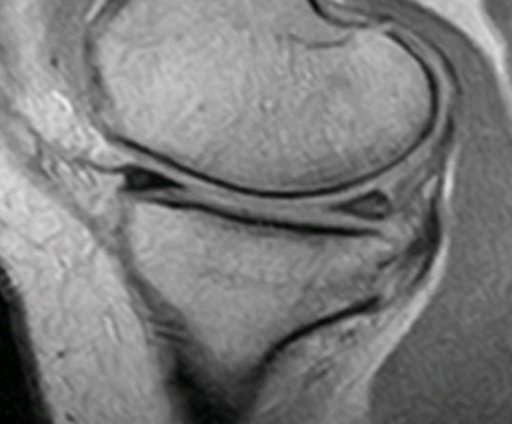
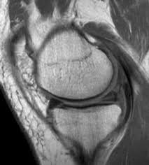
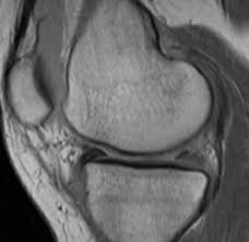
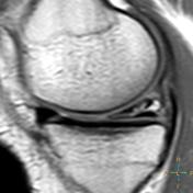
Problems may arise when attempting to distinguish meniscal tears from mucoid intrameniscal change. If proper diagnostic guidelines are followed, there should be little trouble, however, in making this distinction. Normal meniscal tissue is uniformly dark on all imaging sequences. A meniscal tear will manifest as an increased signal within the meniscus on the T-1 weighted images. There may be no signal increase on the T-2 weighted images. It is true that mucoid change will also manifest as an increased intrameniscal signal. A true meniscal tear, however, will feature increased signal that communicates with the surface of the meniscus and usually has more defined margins than mucoid change. Mucoid change does not communicate with an articular surface (1). Figure 1 demonstrates an area of mucoid change in the posterior horn of the meniscus. Compare this appearance with a true tear in figure 2, which is more linear and well defined, and which communicates with the meniscal apex.
Advertisement

Tear Orientations
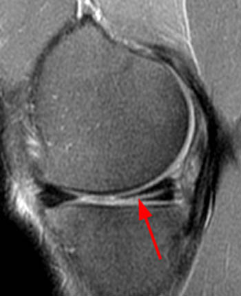
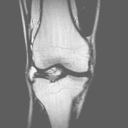
A tear may be of varying size and configuration. The common orientation of tears includes horizontal, oblique and complex. Complex tears manifest multiple lines of tear and are not restricted to a simple linear configuration. Tears resulting from degenerative changes tend to be more horizontal and linearly oriented (fig. 3). Traumatic tears are often obliquely oriented or may exhibit a complex tear pattern.
It is important to note the zone of the meniscus that the tear involves. Is it primarily in the inner, middle or outer third of the meniscus? Tears in the outer, more peripheral third of the meniscus have a chance of healing in some cases, due to a better blood supply. Tears in the inner third of the meniscus have virtually no chance of healing. It is also important to attempt to estimate the length of the tear longitudinally within the meniscus.
Bucket Handle Tear
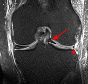
A type of tear that is often discussed, although it is not particularly common, is a bucket handle tear. This type of tear extends longitudinally through the meniscus and is extensive enough that the inner portion of the meniscus becomes detached and flips into the intercondylar region of the joint. This results in a meniscus that appears truncated or too small because part of it has been displaced. In addition, the flipped portion of the meniscus is often visible in the intercondylar region and may end up adjacent to the posterior cruciate ligament (PCL), giving the impression that there are two PCLs on the coronal images. This is a named sign and is termed the “double PCL sign.” Figure 4 demonstrates a bucket handle tear of the lateral meniscus with a small meniscal body and a flap of the meniscus, which is displaced into the intercondylar region.
A region that is often overlooked is the root of the meniscus at the posterior attachment point on the tibial plateau. A diligent search should be made for tears here. In figure 5 there is increased signal involving the posterior root of the meniscus, and it appears somewhat fragmented, representing a tear within the root.
Meniscal Cyst
A tear that communicates with the outer periphery of the meniscus may result in a collection of fluid (2). This is termed a meniscal cyst. These cysts manifest a synovial lining, and, since they contain fluid, appear bright on the T-2 and fat-suppressed images with a dark signal on the T-1 weighted images. Figure 6 demonstrates a small meniscal cyst associated with a complex posterior horn tear. Cysts are often found at the medial aspect of the knee, as demonstrated in figure 7.
Meniscal tears when traumatic in nature are often associated with anterior cruciate ligament and medial collateral ligament tears.
References:
1. Stoller D. W.: Magnetic Resonance Imaging in Orthopaedics and Sports Medicine, ed 2. Philadelphia, J. B. Lippincott Company 1993.
2. Kaplan P. A. et al.: Musculoskeletal MRI, ed 1. Philadelphia, W. B. Saunders, 2001.


