


MRI Appearance in Piriformis Syndrome

Piriformis syndrome (PS) is a diagnosis frequently seen in chiropractic practices. It is a relatively common condition and usually diagnosed based upon clinical findings. It is common, however, to see imaging findings on MRI for this condition. PS may exhibit symptoms similar to lumbar disc herniation in some cases, i.e., chronic gluteal pain with radiation into the lower extremity similar to L5-S1 radiculopathy. In cases where objective corroboration is necessary, MRI could prove to be a useful tool to accomplish this. Other differential considerations that may mimic PS include hamstring muscle injury, sacroiliitis, gluteal muscle strain, ischial tuberosity bursitis, trochanteric bursitis, hip/pelvis stress fractures or intrinsic hip pathology. These are all conditions that could be differentiated with MRI.
PS may affect almost any young to middle age adult; the age range stretches from 18 to 55 and there is a 6:1 predominance for females. Since there are several common causes of piriformis syndrome, the imaging findings may vary somewhat. The most common cause is trauma to the gluteal region, followed by overuse and compensatory contraction due to spinal instability. Less common causes include spinal stenosis, intrinsic piriformis strain, myositis, tumor compression, and prolonged sitting.
Advertisement

Findings on the T-1 weighted images may include hypertrophy (the most common finding), effacement of the fat in the greater sciatic foramen and loss of longitudinal muscle striations. On the T-2 weighted images, in addition to the muscle hypertrophy, there may be diffuse muscle edema with hyperintense signal characteristics, decreased signal characteristics due to fibrosis from trauma, signal change within the gluteus maximus and gluteal atrophy.
The cardinal imaging findings to look for in PS are:
-asymmetry in the size of the piriformis muscles right vs. left.
-signal intensity changes with the piriformis.
Studies performed for the diagnosis of PS should include T-1 and fat-suppressed (STIR) images in both the axial and coronal planes to allow the optimal visualization of the muscle.
MRI Findings
I have included 3 images from 3 different patients to illustrate some typical MRI findings in piriformis syndrome. Figure 1 is a fat-suppressed coronal image. The arrow points to the left piriformis muscle, which exhibits an increased signal intensity when compared to its right-sided counterpart.
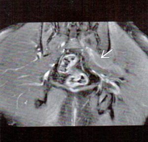
Figure 2 is a T-1 weighted axial image that demonstrates a grossly hypertrophic muscle belly (arrow). This was found in a patient who carried a large wallet in his left hip pocket and sat for long periods of time.
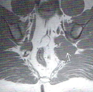
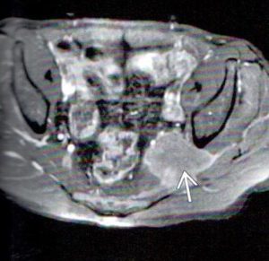
Figure 3 is a fat-suppressed axial image. The left piriformis muscle is both grossly hypertrophic and increased in signal intensity. There is compression and displacement of the sciatic nerve, which is positioned just medial to the posterior aspect of the left ischium.
Treatment
Piriformis syndrome usually responds well to chiropractic care. If a particular case is refractory to conservative care, MRI may be useful to exclude other gluteal pain etiologies, such as those mentioned previously. In extreme cases of prolonged unremitting pain, surgical release of the piriformis or lysis of adhesions/fibrosis surrounding the piriformis or sciatic nerve may be attempted.
References:
1. Stoller D. W., Tirman P. F., Bredella M. A. : Diagnostic Imaging Orthopaedics, ed 1. Salt Lake City, AMIRSYS 2044.


